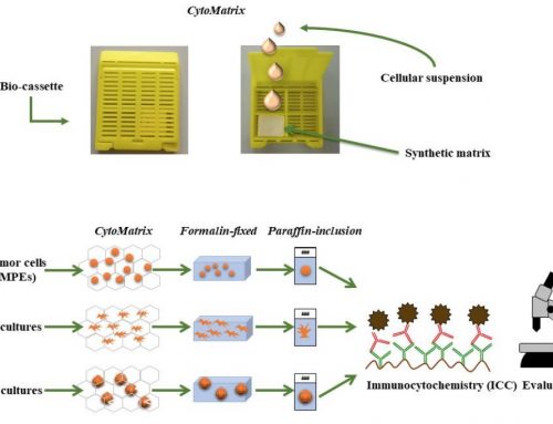
THE APPLICATION OF CYTOMATRIX FOR LEUKEMIC NON-NODAL MANTLE CELL LYMPHOMA DIAGNOSIS
Authors: Antonella Bianchi, Ombretta Annibali, Daniela Righi, Stefano Fratoni, Giuseppe Avvisati, Anna Crescenzi.
A 50-year-old man presented to the Campus Bio-Medico University Hospital for a specialized hematologic examination with incidental lymphocytosis (13.40 x 10³ / uL) and splenomegaly. A CT scan confirms splenomegaly in the absence of lymphadenomegaly. The patient undergoes bone marrow biopsy for suspected lymphoma. Bone marrow histology: Excellent bone marrow tissue with cellularity equal to 95% (hypercellular for the patient’s age) reveals a small lymphoid infiltrate with a diffuse solid pattern equal to about 90% of cellularity with the following immunophenotypic findings: CD20 +, Pax5 +, Cyclin D1 + (weak and partial expression), BRAF + (RM8 BioSB), TdT-, CD10-, bcl6-, CD23-, CD3- and CD5-. There is a small lymphoid component with a diffuse interstitial pattern equal to about 5% of cellularity with the T-cell related phenotypes CD3 + and CD5 +. The CD138 + plasma cell proportion is equal to 3% of cellularity with an IgK: IgL ratio of 1: 1 (unbalanced). Three hematopoietic series in the residual medullary proportion are normally represented and in regular ratios. These findings suggest an indolent peripheral B-cell lymphoproliferative process with an immunophenotypic profile referable to Hairy Cell Leukemia (HCL). We recommend molecular examination and comparison with smears from bone marrow aspirate to confirm the diagnosis.


